

 |
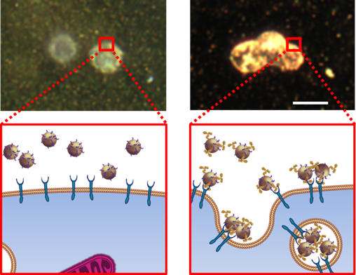
Upper left: A sPA signal cannot
be obtained if the gold nano- particles have no targeting anti- body -- and thus cannot be captured by metastatic cells (drawing bottom left).
Upper right: A strong sPA signal
is obtained from gold nanoparticles with a targeting antibody that are captured by the metastatic cells (drawing bottom right).2 |
CANCER DIGEST – May 22, 2015 – Researchers at the National Institute of Biomedical Imaging and Engineering have developed a highly sensitive and accurate imaging technique for visualizing cancer cells in the lymph nodes.
The non-invasive screening method could one day reduce the need for the current practice of surgically removing lymph nodes to determine whether metastatic cancer cells have invaded the lymph nodes.
The new imaging technique – so far tested only in mice – offers a rapid and effective tool to non-invasively identify very small numbers of these cells, known as micrometastases, thus detecting cancer’s spread at its earliest stages, which is critical for timely treatment.
It works by combining a tiny gold nanoparticle with an antibody that specifically targets the metastatic cancer cells. The antibodies latch onto the micrometastases and the nanoparticles, which are visible to the imaging system, then shows where the cancer has spread.
The work, developed at the University of Texas at Austin and the University of Texas MD Anderson Cancer Center, is reported in the October issue of Cancer Research. The imaging technique is known as ultrasound-guided photoacoustics combined with nano-sensors designed to target and identify metastatic cells in lymph nodes.

No comments:
Post a Comment