

 |
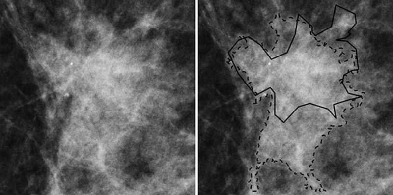
| The 3CB artificial analysis identified the areas of suspected cancer on the mammogram outlined in solid and dashed lines. Photo courtesy RSNA. |
In reality researchers are using artificial intelligence to see patterns that are not readily visible to the radiologist looking for tumors, for example, or to a research scientist looking for genetic patterns that spell a likelihood of XYZ disease.
That’s how scientists are using artificial intelligence to determine which suspicious lesions in the breast need to be biopsied. Led by Karen Drukker, PhD and Maryellen Giger, PhD at the University of Chicago and John Shepherd, PhD at the University of Hawaii their research team has developed a method of analyzing the lipid (fats), protein tissue, and water in lesions to determine if a lesion is just a benign lump of cells or a lesion that needs to be biopsied to determine if it is malignant. Their research appears in the the Dec. 11, 2018 journal Radiology.
Currently about 32 percent of such lumps seen on a mammogram and referred for biopsy turn out to be cancer. Using their artificial intelligence system, they call 3CB, the researchers have been able to raise that percentage by analyzing the lipid, protein and water components of the lesions.
In a recent study the researchers used their AI analysis on mammograms from 109 women with breast masses that were suspicious for malignancy and were referred to biopsy. Visual interpretation alone identified 35 of the masses that turned out to be cancerous, while 74 were benign. Using 3CB analysis of the lesions identified 50 percent of the lesions as being cancerous and missed only one cancerous lesion.
While the method is not ideal at this point, it shows promise of being able to significantly improve radiologists’ ability to identify the lesions that need to be biopsied and ultimately reducing the number of unnecessary biopsies. The researchers next want to use their system in conjunction with breast tomosynthesis or 3D breast imaging to further improve the accuracy of the method.
Source: Radiological Society of North America press release

No comments:
Post a Comment